Showing posts with label Protists. Show all posts

Saturday, September 18, 2021

Sporozoa: Definition, Characteristics, Classification, Reproduction, Examples and Role in Life

Plasmodium malariae"Plasmodium malariae" by Michael Wunderli is licensed under CC BY 2.0

Definition of Sprozoa

The term Sporozoa comes from the Greek, namely  spore  which means "seed" and  zoa  which means "animal". Sporozoa are the only members of Protozoa that do not have locomotion and move by launching their bodies in the medium in which they live. As the name implies, Sporozoa has a characteristic that is able to form spores in one stage of its life cycle.

There are 4,000 species of Sporozoa, most of which live as parasites on animals and humans. The body of Sporozoa is spherical or oval in shape, has a nucleus but lacks contractile vacuoles. The adult form has no means of movement. Many Sporozoans have complex life cycles, in certain phases living on a single host and at other phases living on different hosts.

In their life cycle, Sporozoa show an alternation of offspring between the vegetative and generative phases. Immature sporozoa are called sporocytes  which move easily along the bloodstream. All Sporozoa form thick-walled spores when they are in the zygote stage. These spores are fixed structures that are dispersed through food, water, or insect bites.

Although Sporozoa do not have locomotion, they contain complex organelles that help them attach to and attack the host. Many of its members have complex life cycles. Therefore, the class Sporozoa is also called Apicomplexa. One of the well-known examples of Sporozoa is the cause of malaria, namely  Plamodium .

Characteristics of Sporozoa

Sporozoa or Apicomplexa have several characteristics or characteristics that distinguish them from the other three types of Protozoa . Here, the author describes the characteristics of Sporozoa in general.

■ Do not have a special motion tools, so Sprozoa they move by sliding or varying the position of his body.  

■ It is a single-celled organism (unicellular).  

■ Plumpness parasitic, both animals and humans.  

■ It can form spores at some point in their life cycle.  

■ Have spores oval.  

■ The size of the spores around 8  -  11 microns on a wall of chitin.  

■ Has 2 polar capsules at the anterior, paired with a form like a pumpkin, the same size, located on the corner of the longitudinal axis with the posterior end.  

■ From the front of the anterior end equal to the width of the posterior.  

■ Wall valve unclear.  

The life cycle of Sporozoa shows alternation of generations/offspring between sexual (generative phase) and asexual forms (vegetative phase).  

■ The body is round or oval.  

■ Have a nucleus (center cell) but does not have a contractile vacuole.  

■ Have special complex organelles at one end of the cell (apex) which serves to penetrate cells and tissues of the host.  

■ The process of absorption of food, breathing (respiration) and expenditures (excretion) occurs directly through the body surface.  

■ Most Sporozoa species cause disease in the host (host) the host.  

Sporozoa Classification

The sporozoa class has 3 (three) different characteristics between one genus and another, the differences include:

■ Genus sporozoa who live in red blood cells and require biological vectors, these properties are in Genus  Plasmodi - um .  

■ Genus sporozoa who live in the intestinal and does not require biological vectors, these properties are in the genus  isospora  and Genus  Eimerie .  

■ Parasites that live inside endothelial cells, leukocytes mono - nucleus, body fluids, cells and the host tissue is unknown biological vectors, these properties contained in the genus  Toxoplasma .   

Parasites belonging to the class sporozoa reproduce asexually (schizogony) and sexually (sporogony) alternately. Both of these ways of reproduction can take place in the same host, as occurs in the subclass Coccidia. Meanwhile, what takes place in two different hosts is found in the Haemosporidia subclass (Plasmodium). The sporozoa class can be classified - sikan as shown in the following diagram.

How to Reproduce Sporozoa

The way of reproduction in Sporozoa (ex. Plasmodium) was first discovered by Ronald Ross and Grassi. Reproduction in Sporozoa can occur in two ways, namely:

️   Asexual reproduction (vegetative)  that occurs in the human body  skizogoni  (cleavage in the host's body remains) and on the body of the female Anopheles mosquito is  sporogoni  (sporulation on the host while).

️   Sexual reproduction (generative)  by melting makrogamet and mikrogamet in the body of the Anopheles mosquito.  

Read: Pictures and Stages of the Plasmodium Life Cycle in the Human Body and Mosquitoes.

Examples and Roles of Sporozoa in Life

As explained earlier that Sporozoa are parasitic in both animals and humans and mostly cause disease. Therefore, it is found that there are many detrimental roles compared to the beneficial roles of Sporozoa. Then what is the detrimental role of this Sporozoa? Here are some examples of Sporozoan organisms and their role in life.  

️ ■  Babesia bigemina  is a species of disease-causing Texas fever.

️■   Theileria parva  is a species of disease-causing East Coast fever (Africa).

■ Toxoplasma gondii   is a species Sporozoa cause Toxoplasmosis disease that causes meningitis, hepatitis and fetal infection. These organisms enter the human body through food, for example meat contaminated with toxoplasma cysts from cat or bird feces. Toxoplasma gondii infection is   dangerous for pregnant women because it can cause babies born with mental disabilities, blindness, and swelling of the liver.

■️   Plasmodium vivax  is the cause of malaria tertian. Sporulation period (spore formation period) every 2 x 24 hours.

■️   Plasmodium ovale  is a cause of disease of the spleen. The sporulation period is every 48 hours.

️■   Plasmodium malariae  is a cause of malaria quartana. The sporulation period is every 3 x 24 hours. 

■ Plasmodium falciparum  is the cause of malaria Tropikana. This Plamodium has a sporulation period of about 1 day (1 x 24 hours). 

With so many types of malaria caused by Plasmodium, a drug was developed to prevent the spread of malaria. One of them is the drug  chloroquinone  (quinine) which can kill the malaria parasite. Unfortunately, this parasite has the ability to increase his body's immunity to  chloroquinone .

The Anopheles mosquito extermination program did not run smoothly because these mosquitoes became resistant or resistant to pesticides (anti-pest agents). The researchers hope to use genetic engineering techniques to give the Anopheles mosquito the ability to kill the Plasmodium parasite, not spread it.
Ciliates: Definition, Characteristics, Classification, Reproduction, Examples and Roles in Life

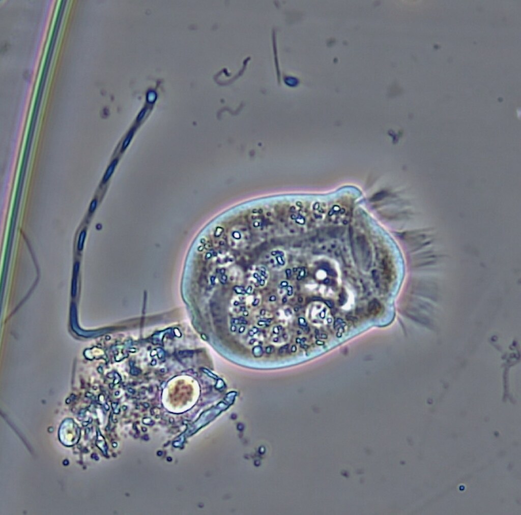
Ciliated Protozoa: Vorticella"Ciliated Protozoa: Vorticella" by bccoer is licensed under CC0 1.0

 Definition of Ciliates

The term "ciliata" comes from the Latin  cilia  which means "little hair". Ciliates are protozoa that have a locomotion tool in the form of  vibrating hairs  (cilia). This vibrating hair is a characteristic of ciliates and functions as a means of locomotion and foraging for food. Ciliates are single-celled organisms (unicellular) with a fixed or unchanged shape.

There are about 8,000 types of ciliates that move with these cilia (shaking hairs) and most of them live in freshwater waters. Ciliates are divided into two groups based on the distribution of cilia, namely cilia on some cells only and cilia that cover all parts of the cell. Food Ciliates are microscopic bacteria and algae. Ciliates get their food by moving the cilia to cause  a whirlpool effect  so that food enters the whirlpool.

Ciliates have many specialized organelles including cilia (singular cilium), short hair-like structures on the outside of their bodies. As previously explained, there are cilia or cilia that cover the entire body surface or are only localized to certain body parts. In the Paramaecium genus , cilia cover the entire body surface.

A good coordination system in hair vibrates, causing Ciliates to move quickly, about  one millimeter per second . Although only single-celled (unicellular), Paramaecium can respond to the surrounding environment well. If it encounters a hazardous chemical or barrier, the cell quickly retreats with cilia moving in a different direction.

Ciliates are excellent predators. Some ciliates, including Paramaecium and Didinium, are able to immobilize their prey by releasing needles called  trichocysts  that attach to their bodies. The prey is then carried into a mouth-like structure and digested in a vacuole which occasionally functions as a stomach.

When the process of digestion of food in ciliates has been completed, the waste products of metabolism will be excreted through  exocytosis . In the ciliate body, excess water will accumulate in the vacuole which periodically (periodically) contracts to empty the fluid through an opening called the  anal pore .

Characteristics of Ciliates

Ciliata or Ciliophora has several characteristics or characteristics that distinguish it from the three other types of Protozoa . Here the author describes the characteristics of Ciliates in general.

■ Moving with cilia or hair shakes.  

■ It is a single-celled organism (unicellular).  

■ Having a permanent body shape or unchanged.  

■ Characteristically heterotrophic, meaning that living with other organisms prey because they can not make their own food.  

■ Generally microscopic, but there are also species with size up to 3 mm so that it can be observed with the naked eye.  

■ Body shape assortment, such as an oval shape, slippers, a bell, a funnel and so forth.  

■ Most live in waters such as swamps, rice paddies and watery places that are rich in organic matter.  

■ Living independently (solitary), parasites, or symbionts in the gut of vertebrates.  

■ Have a contractile vacuole which serves to regulate cell osmotic pressure (osmoregulation).  

■ Has two kinds of nucleus (center) within a single cell, which is  makronukleus  role in metabolism and asexual reproduction (vegetative), and micronucleus  that play a role in sexual reproduction (generative).  

Ciliates Classification

Based on the distribution or distribution pattern of cilia (shaking hair), Ciliates are grouped into two groups, namely Ciliates with cilia that are spread evenly over the entire body surface (ex. Coleps, Bursaria, Paramaecium, Stentor, Calpoda and Prorodon) and Ciliates with localized cilia. or only found in certain body parts (ex. Acineto, Didinium, Stylonichia, and Vorticela). For clarity, please look at the following image.

Meanwhile, based on their way of life, Ciliates are divided into four groups, namely Holotricha, Suctoria, Peritrichia, and Spirotichia. The following is an explanation of each of these ciliate groups.

■ Holotrichia , is a group Ciliate living with swim freely, for example on Paramaecium and Didinium.  

■ Suctoria , is Ciliate group that has tentacles and usually live attached to the substrate, for example Vorticella.  

■ Peritrichia , is Ciliate groups that live in colonies and are usually spherical or oval, for example  Nyctoterus ovalis .  

■ Spirotrichia , is a group Ciliate shaped like a trumpet and a sedentary life in freshwater bergenang or flow, for example, Stentor and Euplotes.  

How to Reproduce Ciliates

Ciliates can reproduce in two ways, namely sexually (mating) through  conjugation  and asexually (not mating) through   transverse binary fission . Conjugation in Ciliates does not produce new daughter cells, but after conjugation, the cell divides to produce four identical daughter cells which are better able to survive the unfavorable environmental conditions.

Binary fission in Ciliates can be observed when one cell divides into 2, then into 4, 8 and so on. This division begins with the splitting of the micronucleus and is followed by the division of the macronucleus. Then 2 daughter cells will be formed after the replacement of the plasma membrane. You need to know that each of the daughter cells is identical and the other cell apparatus has two nuclei and cytoplasm.

Examples and Roles of Ciliates in Life

Similar to Rhizopoda, organisms belonging to the Ciliata class also have various important roles for human life, both harmful and beneficial. Then what is the role of this Ciliata? Here are some examples of Rhizopod organisms and their role in life.

■ Balantidium coli  is a protozoan parasite that lives in the intestines of humans and is the only species that cause disease Siliata. These ciliates can cause sores (inflammation) called  balantidiasis . Balantidiasis is a type of stomach disorder such as bloody diarrhea. The intermediate host of this disease is pigs, then it is transmitted through food or drink contaminated by pig manure containing  Balantidium coli .  

■ Paramaecium  often called animal slippers for cell shape resembles a slipper. It is a single cell organism usually less than 0.25 mm in length. Paramecium has two nuclei, a large nucleus called the macronucleus and two smaller nuclei called the micronucleus. Without macronucleus Paramecium cannot live and without micronucleus Paramecium cannot reproduce.  Reproduction is done asexually by binary fission. Sometimes also reproduce sexually by conjugation. Paramecium is found in abundance in freshwater bodies almost all over the world. Several species of Paramecium are found living in the sea.  Paramecium caudatum  is one type of freshwater Paramecium that is widely used for research.  

■ Nyctoterus ovalis  is a species that lives ciliate parasites in the intestines of cockroaches.  

■ Stentor is shaped like a trumpet Ciliate and settle somewhere.  

■ Vorticella is shaped like a bell Ciliate long-stemmed with straight or spiral shape that comes cilia (hairs vibrate) around his mouth.  

■ Didinium is Ciliate that act as predators in aquatic ecosystems, ie predators Paramaecium. 

■ Stylonichia is Ciliate that looks like a snail or oval, cilianya group called  cirri . Stylonichia is commonly found on the surface of leaves that are submerged in water.

Friday, September 17, 2021

Rhizopods: Definition, Characteristics, Classification, Reproduction, Examples and Role in Life

Amoeba (Amöbe) - Vahlkampfia sp - 630x"Amoeba (Amöbe) - Vahlkampfia sp - 630x" by Picturepest is licensed under CC BY 2.0

Definition of Rhizopods

The term rhizopoda comes from the Greek, namely  rhizo  which means "root" and  podos  which means "foot". Thus, Rhizopoda means feet that resemble roots. Rhizopoda is a Protozoa that has a means of locomotion in the form of pseudo-legs (pseudopodia). Called pseudopodia or pseudopods because they are formed as a result of the protrusion of the cytoplasm of the cell, which seems to function as legs. In addition to moving, pseudopodia also function to find food.

Currently, about 40,000 species of Rhizopoda or Sarcodina are known, namely Protozoa whose shape is not fixed, always changing. One of the most famous examples of Rhizopoda members is the  Amoeba  which can live in fresh water, salt water, in moist soil, and some species live as parasites on animals and humans.

When moving, the  Amoeba  will extend the pseudopodia and hook the ends then release more cytoplasm into the pseudopodia. This kind of motion is called  amoeboid motion . With this pseudo-leg, it means that the shape of Rhizopoda cells changes both at rest and when moving.

Characteristics of Rhizopods

Rhizopoda or Sarcodina has several characteristics or characteristics that distinguish it from the other three types of Protozoa . In the following, the authors describe the characteristics of Rhizopods in general.

■ Moving with false feet (pseudopodia).  

■ Characteristically microscopic, as most have a body size of about 200  -  300 microns.  

■ Having a form of cell that is not fixed alias changeable (ex.  Amoeba ).  

■ Some types have a shell or external skeleton (ex.  Foraminifera  and  Radiolaria ).  

■ Characteristically heterotrophic, meaning it can not make their own food substances so as to meet their nutritional needs, Rhizopoda must prey on other organisms.  

■ Live free (solitary) or parasites.  

■ Swallowing food particles by  phagocytosis .  

■ Breathe by diffusion across the surface of the body.  

■ The cytoplasm consists of ectoplasm and endoplasmic.  

■ Have a cavity in the form of food vacuole to digest food.  

■ Have a contractile vacuole which serves to remove waste products of metabolism and to regulate the osmotic pressure of the body.  

■ Have habitat in fresh water, sea water, wet places and a small part of life in animals or humans.  

■ Some species can form cysts, which form a plasma thickening which serves to protect themselves from unfavorable environment.  

Classification of Rhizopods

Class Rhizopoda or pseudo-legged animals are divided into 5 kinds of orders, namely the order Labosa, order Filosa, order Foraminifera, order Helioza and order Radiolarian. The characteristics or characteristics of each order are as follows.

■ Order Labosa , its features is to have pseudopodia (false feet) short and blunt and can be clearly distinguished between ectoplasm and endoplasmic.  

■ Order Filosa , its features is to have a smooth pseudopodia similar to the yarn and also branched.  

■ Order of Foraminifera , its features is to have a long pseudopodia and also smooth and has a skeleton of lime (calcium carbonate).  

■ Order Helioza , its features is to have the thread-shaped pseudopodia radien and antarfilamen were never united to form the mesh or webbing.  

■ Order Radiozoa , its features is to have a frame made of silica.  

How to Reproduce Rhizopods

Rhizopods (ex.  Amoeba ) reproduce asexually or vegetatively by binary fission. Binary fission in Rhizopods does not go through the stages of mitosis. Cleavage begins with the splitting of the cell nucleus into two, followed by the division of the cytoplasm. The division of the nucleus creates a very deep indentation that will eventually break, resulting in two daughter cells. The two daughter cells will undergo binary fission again so that they become four, eight, sixteen cells and so on. 

In unfavorable conditions, Rhizopods can survive by forming cysts, namely with the inactive body turning into a round shape so that the plasma membrane thickens to protect the body from adverse external conditions. If the external conditions are favorable, for example there is enough food, then the kisat wall will break and the Rhizopoda will come out to start its life again.

Examples and Roles of Rhizopods in Life

Similar to Flagellates, organisms belonging to the Rhizopoda class also have various important roles for human life, both harmful and beneficial. Then what is the role of this Rhizopoda? Here are some examples of Rhizopod organisms and their role in life.
1. Amoeba
Based on their habitat or place of life,  Amoeba is  divided into two genera, namely  Ectoamoeba  and Entamoeba genus  . The following are the differences and examples of species from the two Amoeba  genera  .
A. Ectoamoeba
Ectoamoeba  are amoeba that live freely outside the body of living things. They usually live in humid places. Examples of ectoamoeba  are  Amoeba proteus  and  Chaos carolinese .
B. Entamoeba
Entamoeba  is an amoeba that lives in the body of organisms (animals and humans) and usually causes disease to the host organism it lives on. Examples of  Entamoeba  are as follows.
■ Entamoeba histolytica , the parasite lives in the human intestine and can cause dysentery amoebawi or known by the disease amebiasis . Amebiasis is a disease that causes damage to body tissues, especially erythrocytes (red blood cells) and lymph, causing the patient's face to be mixed with blood and mucus.  
■ Entamoeba ginggivalis , live as parasites in the mouth that can cause inflammation and bleeding gums disease. This amoeba can live in between dirty teeth. In order not to get attacked, brush your teeth after eating and before going to bed.  
■ Entamoeba coli , live in the colon (large intestine) man who is not a parasite but sometimes cause diarrhea (defecate constantly).  

2. Foraminifera
Foraminifera  have shells of organic matter and hard calcium carbonate. Foraminifera live in piles of sand or attached to plankton, algae and rocks. Pseudopodia or pseudo-legs are cytoplasmic strands that function to swim, catch prey and form shells.

About 90% of Foraminifera have been fossilized and their shells are components of oceanic sediments. Foraminifera fossils are   used as  markers of the age of sedimentary rocks and clues in the search for petroleum sources. An example of Foraminifera is  Globigerina .

3. Radiolaria
Radiolaria  live in the sea, the shell is made of silica with different shapes in each species. The dead radiolaria will settle to the bottom of the water as radiolarian mud. Radiolarian mud is used as a scouring agent and explosives. Examples are Colosphaera  and  Acanthometron.

4. Dyflugia, Arcella and Helioza
These three types of Rhizopods live in fresh water.  Diflugia  can produce mucus that causes fine grains of sand to stick together.  Arcella has a shell composed of chitin or phosphoprotein substances. The upper body shell is dome-shaped, while the lower part is concave with holes for the exit of pseudopodia.  Helioza  (solar animals) have pseudopodia that are rigid and shells containing chitin or silica like glass.

Flagellates: Definition, Characteristics, Classification, Reproduction, Examples and Role in Life

Flagellat - 1000x - 41µ"Flagellat - 1000x - 41µ" by Picturepest is licensed under CC BY 2.0

Definition of Flagellates (Mastigophora)

Flagellata comes from the word  flagellum  which means "feather whip". Flagellates are also often referred to as Mastigophora. The word "mastigophora" comes from the Greek  words mastig  which means "whip" and  phoros  which means "movement". Thus it can be concluded that Flagellates are a type of Protozoa that has a locomotion tool in the form of whip feathers (flagellates).

Flagellates are the ancestors of animals and plants. Flagella in Flagellata are located at the anterior end of the body. In addition to functioning as a locomotion, flagella can also be used to determine the state of the environment and collect food by producing a flow of water around the mouth so that food can enter the mouth. Flagellate cytoplasm is surrounded by a pellicle or a clear sheath that gives it a fixed body shape.

Flagellate Characteristics

Flagellates or Mastigophora have several characteristics or characteristics that distinguish them from the other three types of Protozoa . In the following, the authors describe the characteristics of flagellates in general.

■ Moving with feather whip (flagellum).  

■ Live free (solitary), saprophyte, parasites in animals and humans, and there is also a symbiotic mutualism.  

■ Have a pellicle which is a flexible membrane to protect and give shape remains even without the outer frame structure.  

■ Characteristically microscopic, mostly flagellates can not be observed by the naked eye (without the aid of a microscope ).  

■ generally have an oval body shape, long and round.  

■ Characteristically if live solitary unicellular or multicellular if live in colonies.   

■ There are mitochondria and some are not.  

■ Unable to form a cyst.  

■ Habitat flagellates mostly fresh water, sea water, wet ground or in the living body as a parasite.  

Classification of Flagellates

Judging from the shape of the body, flagellates are divided into two types, namely flagellates shaped like plants called  phytoflagellates  and flagellates shaped like animals called  zooflagellates . The differences between the two types of flagellates are as follows.

Phytoflagellates

Phytoflagellates  are flagellates that have plastids and chlorophyll pigments that are used to carry out the photosynthesis process so that  phytoflagellates are autotrophs. In the aquatic environment,  phytoflagellates  act as  phytoplankton  that supply food for other organisms. Based on the shape of the body and the number of flagella it has,  phytoflagellates  are grouped into three classes, namely:

1) Euglenoids

Euglenoids have a body shape that resembles a spindle and is covered by a pellicle. Euglenoids have one or two flagella at the anterior end. At the anterior end there is a red eye spot that contains the pigment carotene. These eye spots serve to protect the light-sensitive area at the base of the flagella.

The best known member of the Euglenoida group is  Euglena viridis.  Euglena viridis is  commonly found in fresh water with the following characteristics:  

■ It has a body size of 35-60 microns   

■ The tip of a tapered body with a feather whip, so it can move on with flagella. This movement is also known as euglenoid motion .   

■ Have a stigma (red eye spots) to distinguish between dark and light  

■ Having chloroplast containing chlorophyll used for berfotosintetis. There are also  non-chloroplast Euglena  , such as  Astasia .   

■ Food entry through sitofaring leading to the vacuole and in the vacuole of food in the form of tiny organisms will be digested.   

2) Dinoflagellates

Dinoflagellates vary in body shape but are mostly oblong with brownish and yellowish coloration. Dinoflagellates are constituents of marine plankton. Although most of them live in the sea, but some live in fresh water. Dinoflagellates are symbiotic in coral reefs, jellyfish, anemones and other invertebrates. The flagella are located in a transverse depression that surrounds the body.

Many species of dinoflagellates lose their flagella and grow as a non-motile vegetative phase. Examples of dinoflagellate members include  Ceratilum, Noctiluca milliaris,  and  Gymnodinium .  Noctiluca milliaris  mostly lives in seawater and has the following characteristics.

■ It has two flagella that is one long and one short   

■ Make a symbiosis with certain types of algae 

■ The body can emit beams exposed mechanical stimulation. We can see it at night, when the waves break the rocks or the paddles hit the sea water, there will be a sparkling light produced by  Noctiluca . This event is known as  bioluminescence .  

3) Volvocida

Volvocida are generally spherical in shape and live solitary or in colonies. Volvocida has 2 flagella. The cell wall of Volvocida is composed of cellulose. For example, the most famous member of this group is the  Volvox globator . The characteristics of volvox are as follows.

The colony consists of thousands of single-celled individuals and each has two flagella.  

■ Each cell has a nucleus, contractile vacuole, stigma, and chloroplasts.   

■ The cells are connected by threads of protoplasm that form a physiological relationship.  

Zooflagellates

Zooflagellates  are flagellates that do not have chlorophyll pigment and are heterotrophs.  Some zooflagellates  are free-living but most are parasites with animal-like shapes. Some of the best known examples of  zooflagellates are  those of the species of the genera  Trypanosoma and  Leishmania . The following is a complete description of the two genera.

1) Trypanosoma 

Trypanosoma have long flat bodies like leaves and do not form cysts. Trypanosomes live in the red blood cells, white blood cells and liver cells of their vertebrate hosts. Infection due to Trypanosoma is also known as  trypanosomiasis . In its life cycle, Trypanosoma has two forms, namely flagellated in the extracellular phase and unflagellated in the intracellular phase. Part of its life cycle is attached to gastric cells or sucks human blood. Intermediate hosts (intermediaries) Trypanosoma are blood-sucking animals such as rat fleas,  Tabanus flies, tse-tse flies,  Glossina palpalis  flies and Glossina morsitans flies  .  

Examples of the types of Trypanosoma are as follows.

■ Trypanosoma lewisi,  living in mice, the intermediary host is a rodent infestation.  

■ Trypanosoma evansi , causes disease sura (lazy) in cattle, the intermediary host is tabanus flies.   

■ Trapanosoma brucei,  causes nagano disease in cattle, the intermediary host is the tse-tse flies.  

■ Trypanosoma gambiense  and  Trypanosoma rhodesiense . Animals that cause sleep in humans were originally found in Africa, then spread to Asia. The intermediate hosts were the fly  Glossina palpalis  for  T. gambiense  and the fly  Glossina mursitans  for  T. rhodesiense .   

■ Trypanosoma cruzi,  the cause of anemia in children (cagas). Trypanosoma Cruzi is  found in Central America.

2) Leishmania 

Leishmania is a cause of disease in the endothelial cells of blood vessels. Endothelium is an epithelial cell that lines the heart, blood vessels and lymph vessels. Examples of types of Leishmania are as follows.  

■ Leishmania donovani,  causes kala azar disease characterized by fever and anemia. This species is found in Egypt, around the Mediterranean and India.   

■ Leishmania tropica,  cause a skin disease called oriental disease. This species is found in Asia (Mediterranean area) and parts of South America.  

■ Leishmania brasilliensis ,  cause skin disease in Mexico and Central America and South America.   

How to Reproduction Flagellates

For flagellates  , phytoflagellates reproduce in two ways, namely sexually by conjugation and asexually by dividing. As for the zooflagellate type flagellates  , reproduction occurs asexually by longitudinal binary fission, while sexual reproduction is not widely known.

Examples and Roles of Flagellates in Life

It has been explained previously that many flagellates live freely in moist soil, water, symbiotic habitats, live inside other organisms with mutualism or parasitic relationships. Here are some examples of flagellate organisms and their role in life.

■ Trichonympha  and  Myxotricha  

2 This type of flagellates lives in the intestines of termites which helps termites to digest wood because they can secrete cellulose enzymes. This enzyme makes the wood particles softer, so they are easily broken down and broken down into small pieces and then absorbed by termites. This absorbed material is partly needed by termites and partly for the survival of Flagellates. Thus intertwined symbiotic mutualism (mutual benefit).

■ Trichomonas vaginalis  

When viewed from the name, this type causes one type of vaginitis, which is an inflammation of the vagina which is characterized by discharge and accompanied by a burning sensation and itching. This species does not have a cyst stage and spreads as a venereal disease. Can also infect and spread in men causing prostatitis.  Trichomonas vaginalis  can be passed from woman to man through sexual intercourse.

■ Giardia lamblia  

Is the only intestinal Protozoa that causes dysentery / diarrhea and cramps in the stomach. These protozoa are found in the duodenum / duodenum. Transmission is through contaminated food or drink and through hand-to-mouth contact.

Euglenophyta: Definition, Characteristics, Classification, Reproduction, Cell Structure, Examples and Role for Life

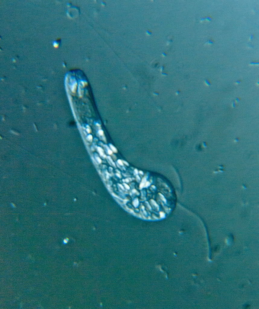
Euglena mutabilis - 400x - 1"Euglena mutabilis - 400x - 1" by Picturepest is licensed under CC BY 2.0

Definition of Euglenophyta

Have you seen plankton? One of the constituents of plankton is the Euglenophyta group, has a single cell (unicellular), and has a true nucleus. Euglenophyta or Euglenoid comes from the Greek, namely  eu  which means true and  gleen  which means eye. Named Euglenophyta because organisms belonging to this group have red eye spots (stigma) that can catch light ( photoreceptive eyespot ) and chloroplasts.

Most of the Euglenoids are autotrophs because they can photosynthesize, some are heterotrophs. The most prominent feature of these protists is their unicellular (single-celled) body, bright green in color and very beautiful. The shape of the euglenoid cell is oval with a more slender posterior part. Although Euglenoids are grouped in the group of plant-like Protists, these living things do not have cell walls like plants. In the absence of a cell wall, they are free to move, so they are often mistaken for animal cells.

Euglenophyta is a unique group of protists because it has properties similar to plants and animals. Considered similar to plants because it has chlorophyll a and b, carotenoids are also found so that he will photosynthesize. Euglenophyta are considered animal-like because they can move actively with the help of one or more whip hairs (flagellates) that come out of their cells. Because it has a locomotion, it can live in waters, such as fresh water and stagnant water.

The results of photosynthesis in Euglenophyta are stored as food reserves in the form of paramylon polysaccharides  . Euglenophyta live as photoautotroph organisms through photosynthesis. However, if the conditions are less favorable, for example there is no sunlight, then Euglenophyta can also live as heterotrophs, by eating the remains of organic matter. Euglenophyta have habitats in fresh water, such as pond water, rice fields, lakes, and are often found in farm ditches which contain a lot of animal waste.

Euglenophyta Body Structure

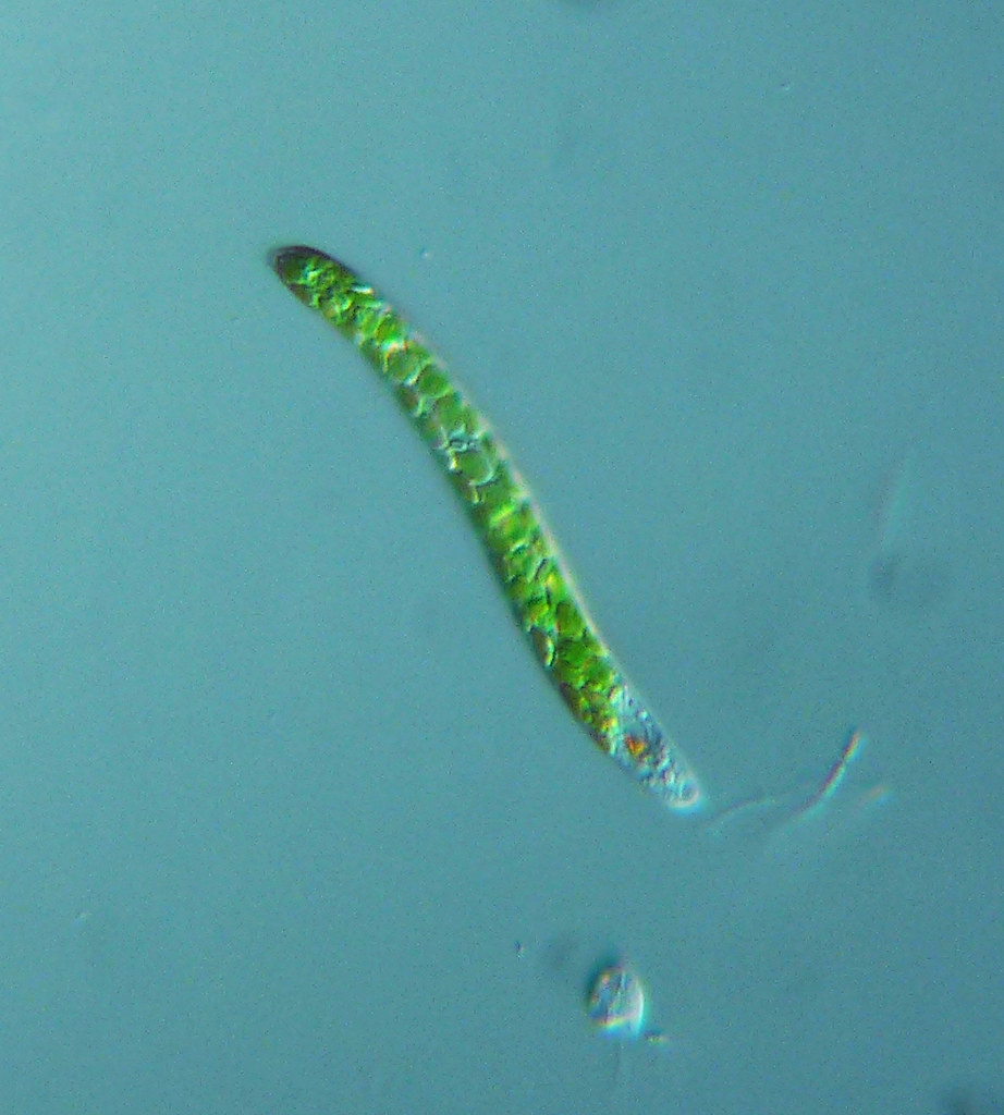
To date, around 1,000 species of Euglenophyta have been identified. One well-known species is  Euglena viridis . Using a light microscope,  Euglena viridis  appears green. Chlorophyll is stored in oval shaped chloroplasts. Euglena is a distinctive member of this group, numbering about 400 species. 

Euglena cells are elongated oval in shape, not rigid, do not have a cell wall containing cellulose, but have a supporting layer of cell membranes and proteins in the form of a flexible pellicle (flexible ) . Thus, he can change shape easily. At one end there is a mouth of the cell and from the mouth of the cell grows several flagella with different sizes. Long flagella are used for locomotion and other flagella are short. Euglenophyta show phototaxis motion, which is the movement of moving places towards the sun.

There is also an eye spot called the stigma. Stigma contains photoreceptors that are covered by red pigment and function to distinguish light and dark. Euglena also has an anterior esophagus, although it is not used for swallowing particulate food. Inside the cell there is also a contractile vacuole whose function is the same as Protozoa.

How does Euglena get her food? These organisms carry out photosynthesis in chloroplasts and are facultative autotrophic. Most of these organisms are able to assimilate organic substances during photosynthesis. In fact, some types of Euglena can swallow food in the form of particles through temporary openings adjacent to the esophagus.

Euglenophyta Characteristics

Euglenophyta or Euglenoids have general characteristics or characteristics, which are as follows.

■ Unicellular (single-celled)  

■ From the mouth appear one to four flagella (whip feathers) which serves as a means of motion.  

■ In general, have flagella of unequal length (Heterokontae)  

■ Characteristically motile (likely to move).  

■ colored green because they contain chlorophyll.  

■ Cells elongated oval.  

■ At one end there is the mouth of the cell.  

■ Generally live in fresh water rich in organic matter.  

■ Autotrophs and or heterotrophic. It is autotrophic, because it has chlorophyll a and b, beta carotene and some xanthophylls. Heterotrophs because they eat organic particles (ex. bacteria ) that are available. Some types of Euglena which are autotrophs can become heterotrophs when light levels are low.  

■ Some have chloroplasts (for photosynthesis) and some are not.  

They are phototrophic (make their own food), osmotrophic (eat by diffusion), and phagotrophic (eat by capturing food).  

■ Backup paramilum food form, that is another form of polysaccharides.  

■ Do not have a cell wall made of cellulose but have thin cell membranes are composed of layers of spiral-shaped proteins.  

■ Have eye spots called stigma. Stigma ( eyespot ) is bright red that is sensitive to light. The red color on this stigma is the pigment astaxanthin. These eye spots serve to protect the light detectors located near the base of the flagella. With the detector, Euglena can move towards the direction of light of the appropriate intensity.  

■ The body is covered pellicle.  

■ Have a contractile vacuole and the food vacuole.  

■ The anterior end of the cell in the form and in the form of lower sitostom esophagus.  

Euglenophyta Classification

Phylum Euglenophyta is divided into three orders, namely:

1. Euglenales

2. Paranemales/Eutreptiales

3. Rhadbdomonadales

Euglenophyta Reproduction Method

How also do Euglenophyta (ex. Euglena) reproduce? Basically the way Euglena reproduces is the same as Protozoa, namely asexually. In general, this group reproduces asexually by longitudinal binary fission. At first it divides according to the longitude axis. The cells have 2 whip feathers and goblet-shaped chloroplasts and contain pyrenoids.

Before splitting, the pyrenoids extend across and the two whip feathers are far apart from each other. The pyrenoids and chloroplasts then make indentations and the cells will divide into two new individuals, each with a whip feather accompanied by the formation of a stigma.

Biologists have observed that Euglenoid reproduction occurs by mitosis, but they have not found sexual reproduction. Euglenoids often divide rapidly, so that chloroplast division has not yet had time to occur. This causes a new individual whose division results do not have chloroplasts and lose their color.

This new individual then grows into a living being that is heterotroph. The nature of the euglenoid, which is sometimes plant-like and sometimes animal-like, causes the grouping of Euglenoids to be a matter of debate.

Examples and Roles of Euglenophyta in Life

An example of a species of Euglenophyta is Euglena (green). Euglena includes all members of the Euglenophyceae whose cells during their lifetime have falgel and can move (motile). Solitary life, never forming a colony. Chloroplasts are disc-shaped and some are ribbon-shaped. Euglena's diet is very varied and includes all living organisms.

If Euglena grows in a dark place with a suitable organic substrate, the color will be lost so that it will be heterotrophic. However, if there is light, then Euglena will be colored again. Euglena's food reserves are in the form of paramylum, which is carbohydrates in the form of ring, rod or round discs, sometimes relatively large in size.

The role of Euglenophyta in life, among others, is as follows.

■ Used as an indicator of water pollution. For example, the surface of the water in which there is a lot of  Euglena viridis , will appear greenish in color. While there is a lot of  Euglena sanguinea  looks reddish.  

■ In the field of fisheries, Euglenophyta is phytoplankton that serves as a food fish.  

■ In the economic field waters, Euglenophyta the primary producers in aquatic ecosystems, namely as a provider of organic matter and oxygen for aquatic animals such as fish, shrimp and aquatic insects.  

■ In the field of science, Euglena is often used as an object of study observation. Because this type of algae is easy to get and breed and as an organic digestive.  

■ In addition to the benefits of unisex loss (negative impact) that ditumbulkan by Euglenophyta, which pollute water sources and leads to accumulation of soil sediment at the bottom of a pond or lake.

Chrysophyta (Golden Algae): Definition, Characteristics, Classification, Reproduction, Examples and Role for Life

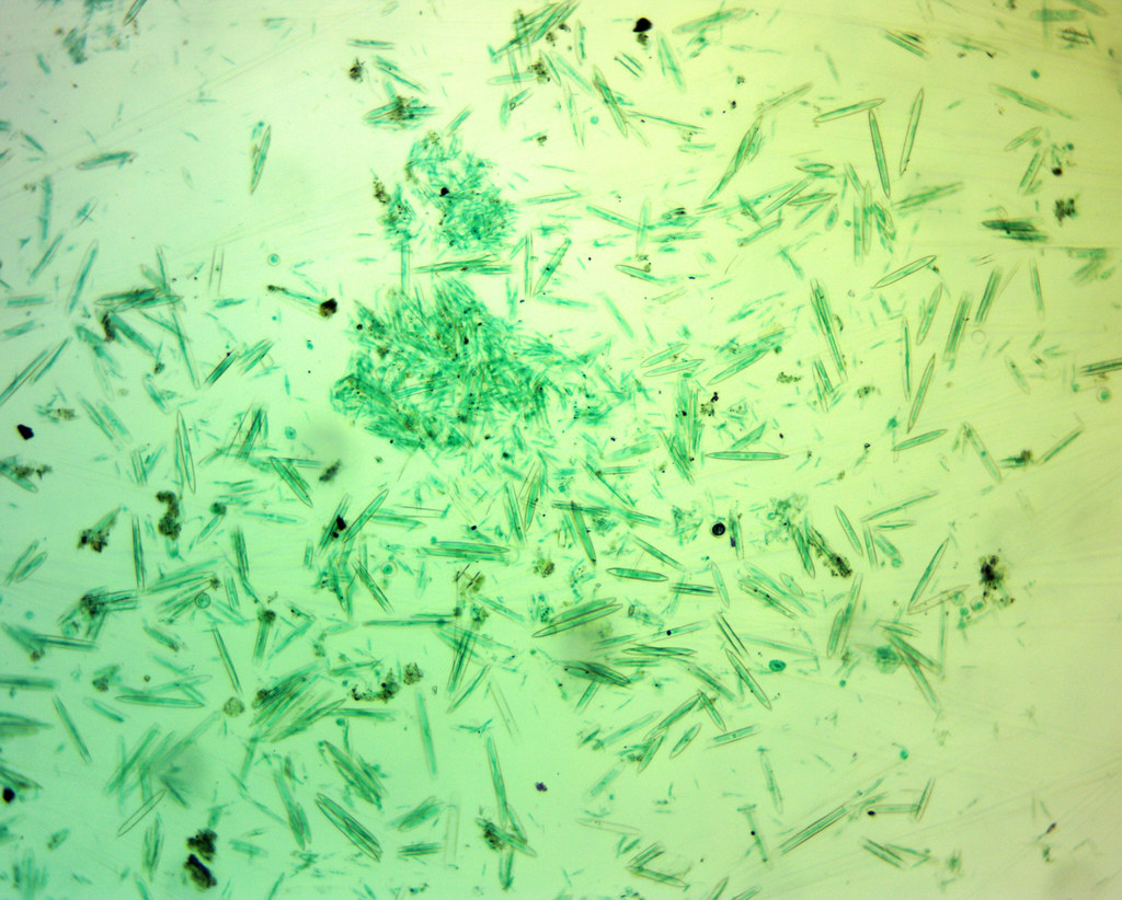
Navicula oblonga - 160x - Schrägsicht"Navicula oblonga - 160x - Schrägsicht" by Picturepest is licensed under CC BY 2.0

Definition of Chrysophyta (Golden Algae)

Chrysophyta algae are also called golden algae ( golden algae ) or blonde algae. The term "Chrysophyta" comes from the Greek,  chrysos which means "golden". The golden color is due to the presence of pigments in the form of carotenes and xanthophylls which are dominant compared to chlorophyll la and c, thus making the plastid cells yellowish green/golden brown. Other sources say that the golden color is caused by a pigment called fucoxanthin ( fucoxanthin ).  

Chrysophyta mostly live in fresh water, although some species exist in sea water. Algae of this group have food stored as laminarin, which is a polysaccharide as food storage in these algae. Golden algae have a variety of structures and shapes. Some do not have cell walls and can crawl like  Amoeba . Some have cell walls made of cellulose.

Most groups of golden algae are unicellular but some form colonies. Blond algae cells have two flagella, so they are called biflagellates, especially for algae whose cell wall structure is composed of pectin. The two flagella are attached near one end of the cell. In addition to living in water, there are also Chrysophyta that live on land.

Blond algae that live on land are often found as velvety membranes at the edges of ponds, waterfronts, or in moist soil. In addition to laminarin, Chrysophyta store excess food in the form of oil so that it is an important component in the formation of petroleum. Phylum Chrysophyta consists of about 5,300 species, and 5,000 of them are diatoms which are now included in a separate phylum, Bacillariophyta.

Characteristics of Chrysophyta (Golden Algae)

Golden algae (Chrysophyta) have the following characteristics or general characteristics.

■ Core are eukaryotic cells because the cell nucleus already had membranes.  

■ There are unicellular (single-celled) and those that are multicellular (multicellular). The unicellular algae in the waters act as a component of phytoplankton.  

■ Characteristically autotorof, because chlorophyll to perform photosynthesis. However, some are heterotrophs by absorbing food.  

■ Habitat in waters such as freshwater, brackish water and seawater, and there is also a land of life, especially in places that are wet.  

■ Anyone have a cell wall and there is not.  

■ The cell walls contain cellulose, pectin or silica.  

■ Most Chrysophyta have flagella to move mainly have a cell wall. But there are also amoeboid (moving crawling like  Amoeba ) for Chrysophyta that do not have cell walls.  

■ Have a pigment carotene, xantofil, chlorophyll a and chlorophyll c.  

■ Most are microscopic (can not be observed with the naked eye).  

■ Live solitary or in colonies.  

■ Keep a spare makana in the form of laminarin or oil.  

Classification of Chrysophyta (Golden Algae)

Golden algae are classified into three classes, namely:

■ Xanthophyceae  (yellow-green algae). Has chlorophyll, xanthophyll. Example:  Vaucheria sp .  

■ Chrysophyceae  (golden-brown algae). Has chlorophyll and carotene. Example:  Ochromonas, Synura .  

■ Bacillariophyceae  (diatoms). Often found on the surface of wet soil (rice fields, sewers, ditches). Unicellular body, some are colonies. The cell wall is composed of two parts, namely the box (hypotheca) and the lid (epiteka). Example:  Navicula ,  Pinnularia . But now diatoms have been separated from Phylum Chrysophyta and included in a separate phylum, namely Bacillariophyta.  

Based on the type of cell, Chrysophyta can be divided into two types, namely:

Unicellular Chrysophya (Single-Celled)

■ Ochromonas , is a type of unicellular Chrysophyta having two flagella, one long and one short. Ochromonas can grow autotrophs by using sunlight energy or heterotrophs by absorbing food.  

■ Navicula , often referred to as diatomaceous or algae grit, boxes or elliptical in shape, if the dead fossils will form diatomaceous earth that serves as an abrasive, mix cement or absorbent nitroglycerine on explosives. Reproduction divides itself by separating the body parts which consist of the hypotheca (box) and the epiteka (lid).  

■ Pinnularia , like diatomaceous. 

Chrysophya Multicellular (Multi-Celled)

■ Vaucheria , live in colonies in the tubular filament that sometimes branched. Types that live on land attached to the surface with rhizoids, namely branches resembling roots that are colorless. Vaucheria filaments are   multinucleated and are not bounded by septal walls called senocytes. In the cytoplasm there is a large vacuole in the center of the cell. In the cytoplasm there are many nuclei, disc-shaped plastids without pyrenoids. Food reserves in the form of oil in the form of oil drops.

How to Reproduce Chrysophyta (Golden Algae)

How do golden algae reproduce? This golden algae can reproduce asexually (vegetatively) and also sexually (generatively). The following is an explanation of the two types of reproduction.

Reproduction Asexually (Vegetatively)

Asexual reproduction is carried out by the  formation of large  multinucleated zoospores that have many flagella as in  Vaucheria . These zoospores are considered as a compound structure consisting of a collection of small zoospores with two flagella, each of which does not separate. After these zoospores are released, they then move with their flagella to a new place. Once settled, the flagella are released and germinate to form   new Vaucheria . In addition to the formation of zoospores, there are also Chrysphyta species which reproduce asexually by dividing as in  Ochromonas .

Sexual Reproduction (Generative)

Sexual reproduction in Chrysophytes is by  oogamy , namely by forming oogonia (forming female gametes) and antheridia (forming male gametes) on the same filament. The resulting egg cell is large with one nucleus containing chlorophyll. Sperm produced by antheridia have small flagella. After fertilization occurs, a zygote is formed. Once released from the parent, the zygote is ready to grow to form a new filament.

Examples and Roles of Chrysophyta (Golden Algae) in Life

In human life, golden algae have many benefits, especially  Navicula  and  Vaucheria .  Navicula  that has died and settles on the seabed forms soil deposits that are useful as abrasives, dynamite insulators, materials for making paints, varnishes, basic materials for the glass industry, filters and vinyl records. In  Vaucheria , food reserves are stored in the form of oil, so this organism is a major component in the formation of petroleum.

Pyrrophyta (Fire Algae / Dinoflagellates): Definition, Characteristics, Cell Structure, Classification, Reproduction, Examples and Role for Life

Blue tide of dinoflagellates"Blue tide of dinoflagellates" by BMC Ecology is licensed under CC BY 2.0

Definition of Pyrrophyta (Fire Algae / Dinoflagellates)

The phylum Pyrrophyta is often called the Dinoflagellates, so named because of the movement of the two whip-like flagella (in Latin, dino  means whirlpool). Dinoflagellates consist of about 1,100 species, mainly living in sea water, although some species live in fresh water. Dinoflagellates are motile unicellular algae, with the main characteristic of having gaps and grooves on the outside of the envelope that surrounds the cell wall.

Some types of dinoflagellates do not have a cell wall, but most have a cell wall that is divided into polygonal cellulose plates that are closely connected to each other. Pyrrophyta are also often called fire plants or fire algae.

Why are Pyrrophyta or Dinoflagellates called fire algae?

Phylum Pyrrophyta is called fire algae because it has a shell that contains phosphorus which is able to emit a bright red light like fire or blue green which is very beautiful, especially in dark conditions at night in sea water. This phenomenon of light fluorescence is called  bioluminescence . An example is  Noctiluca sp. , and  Ceratium sp.

The appearance of red color is because these protists contain a lot of  carotenoids , so their appearance is more often gold, brown or red than green. Pyrrophyta or Dinoflagellates mostly have non-contractile vacuoles, chloroplasts, and have chlorophyll a and b.

Dinoflagellate green chlorophyll is usually covered by a red pigment that helps capture light energy. When the water is warm and rich in nutrients, the Dinoflagellate population will explode. The number of dinoflagellates will be so large that the water will be colored red by body color. This event is known as  a red wave  ( red tide ).

Autotrophic dinoflagellates are a common type of phytoplankton. They are excellent producers of biomass and oxygen. Some dinoflagellates which are photosynthetic, live in symbiosis on the bodies of several types of corals, sea anemones, flatworms, and giant clams.

Some dinoflagellates are also heterotrophs. They live by ingesting organic matter and other living cells. In addition, a small proportion of Dinoflagellates can be parasitic on the bodies of various marine animals, for example  Protogonyaulax catenella .

Pyrrophyta (Fire Algae / Dinoflagellates) Body Structure

To understand the parts of the cell structure of Dinoflagellates and their functions, please look at the pictures and explanations below.

■ Dinoflagellata is essentially motile unicellular organisms and berflagel two (biflagella), golden-brown colored, and including photosynthetic protists. Although the dominant color is golden brown, there are also yellow, green, brown and even blue ones. Some of them are non-motile, do not have flagella, are ameboid, and are fibrous.  

■ cells are generally covered by a coat or a rigid plate made of cellulose arranged artistically sculpted. This arrangement of plates is called  armor plate  or armor plate .  

■ dinoflagellates have two slits or grooves are longitudinal grooves (longitudinal) called the  sulcus  (groove) and a circular groove (transverse) known as the  cingulum  or annulus or corset.  

■ Two flagella in different Dinoflagellata (heterokonts), the flagellum transverse and longitudinal flagellum. Longitudinal flagella are smaller and smoother and point posteriorly and are located in the  sulcus . While the transverse flagella is shaped like a ribbon and is located on the  cingulum . These two types of flagella move in different directions, resulting in a whirlpool when the Dinoflagellates move.  

The nucleus is large and named mesokaryon by Dodge (1966). This part of the chromosome lacks histones or RNA.  

■ plastids or chromatophore have chlorophyll a and chlorophyll c.  

■ vesicles located at the bottom of the cell memberan.  

■ vacuole non-contractile called  pusule  located near the base of flagella.  This pustule is useful for floating on the water surface and osmoregulation. There are no contractile vacuoles in Dinoflagellates.

Characteristics of Pyrrophyta (Fire Algae / Dinoflagellates)

Pyrrophyta or Dinoflagellates or Fire Algae have general characteristics or characteristics, which are as follows.

■ Unicellular (single-celled)  

■ Characteristically motile (active)  

■ It has a flagellum (feather whip)  

■ Have a real cell wall composed of plates that contain cellulose, but there are some who do not have cell walls, for example  Gymnodinium sp.  

■ Have a cell with a characteristic that is there are gaps and grooves as well as in the cells are the plastids containing pigment chlorophyll a and c, as well as carotenoids that the yellowish brown in color.  

■ Characteristically autotrophic (able to perform photosynthesis or are photosynthetic) and serves as phytoplankton in the ocean.  

■ Characteristically are heterotrophic life by way of ingesting organic matter and other living cells.  

■ There are also nature as a parasite that lives in a way attached to the body of a variety of marine animals, for example  Protogonyaulax catenella .  

■ Live free or symbiosis on the body, some corals, sea anemones, flatworms, and shellfish raksaksa.  

■ In some species, cangkagnya containing phosphorus that fluoresces at night.  

■ Most berhabitat in seawater but those that live in fresh water. 

■ Have non-contractile vacuole which serves to float and osmoregulation.  

Classification of Pyrrophyta (Fire Algae / Dinoflagellates)

Because dinoflagellates can be seen as both plant-like and animal-like, their classification has been debated among botanists, zoologists, and paleontologists. The most widely accepted classification scheme is that all dinoflagellates are members of the kingdom Protista, the division Dinophyta, and the class Dinophyceae.

Dinoflagellates were then included in the group of algae (plant-like protists) namely the phylum Pyrrophyta and classified into many orders, genera, and species based on characteristics such as feeding behavior, composition of their outer plate, overall anatomy and physiology.

Pyrrophyta (Fire Algae / Dinoflagellates) Reproduction Methods

Like Euglenophyta , Pyrrophyta also reproduce only asexually, namely by dividing, but some types can produce cysts (resting stage) which are sexual. The cyst will then germinate to produce a new individual under suitable conditions.

Examples of Pyrrophyta or Dinoflagellate species

Dinoflagellates consist of about 1,100 species, mainly living in sea water, although some species live in fresh water. Examples of the most common Dinoflagellate species are  Pfiesteria piscicidia, Gonyaulax catanella,  and  Noctiluca scintillans.  The following is an explanation of the three types of Dinoflagellates.

■ Pfiesteria piscicidia  is a dinoflagellate species are often found off the coast of North Carolina. Scientists have concluded that it is responsible for killing large numbers of fish by secreting poison. This species has an interesting feeding strategy. It is known to use poison to kill fish then wait for it to consume tissue that sloughs off from decaying organisms. This makes it one of the heterotrophic species of several dinoflagellates. 

■ Gonyaulax catanella  is dinoflagellate revolving when they move to use two of their flagella. They are also one of the well-known luminous species of dinoflagellates, as they give off a blue-green glow in the waters they inhabit.  

■ Noctiluca scintillans  are heterotrophic dinoflagellate species that feed on plankton found in estuaries and shallow areas of the continental shelf. This species is often referred to as marine sparkle because it exhibits bioluminescence and becomes very bright when disturbed in water.  

The Role of Pyrrophyta or Dinoflagellates

Dinoflagellates often cause an interesting phenomenon in the sea, which can produce a sudden red color of the sea. This phenomenon is often called  tidal / wave red  or " red tides ". These conditions contain a toxin produced by certain dinoflagellates and can poison fish, shellfish, and sometimes humans.

Toxic red tides can usually occur after the population density of certain dinoflagellates increases sharply ( blooming ). Types of Dinoflagellates that can produce toxic red tides, including Gymnodinium and Protogonyaulax. The toxins or poisons produced by these species are usually neurotoxins or neurotoxins, or can cause the rupture of red blood cells.

When a red tide occurs, thousands of fish suffocate as a result of their gills being clogged or deprived of oxygen by billions of dead and decaying dinoflagellates. However, oysters and mussels "feast" by filtering millions of their food in the water. In this process, their bodies will collect the neurotoxins produced by Dinoflagellates in large enough quantities.

In this situation, Dinoflagellate toxins can accumulate in the body of oysters or mussels without causing the death of the animal. However, if the mollusk is eaten by humans, poisoning can occur in humans who eat it. Therefore, the consumption of shellfish is often avoided during the summer, which is the season when the population of Dinoflagellates increases sharply. 

Thursday, September 16, 2021

Bacillariophyta (Diatoms): Definition, Characteristics, Cell Structure, Classification, Reproduction, Examples and Role for Life

Diatoms"Diatoms" by kevin dooley is licensed under CC BY 2.0

 Definition of Bacillariophyta (Diatoms)

Bacillariophyta or Diatoms are unicellular algae that are widely distributed in freshwater and seawater, as well as in moist soils. The number of diatoms is very large, it is estimated that there are 16,000 types. Due to the large number of diatoms, which act as one of the phytoplankton, they become an important producer component in marine waters.

Some diatoms live alone and some form colonies in filaments. Some live freely on the surface of the water, some other species live attached to the substrate. Bacillariophyta has food that is stored as leukocin in the form of oil drops and has photosynthetic pigments, namely chlorophyll a, chlorophyll c, xanthophyll, and carotene.

Diatom cells elongated shape with a cell wall or shell consisting of two parts like a box (hypotheka) with a lid (epiteka). The shell is composed of pectin and silica with various forms of ornamentation. When a diatom dies, a translucent silica shell remains. The shells of diatoms are equipped with tiny holes that allow the cell to come into contact with the water environment.

Diatoms have the highest abundance and can be found in various types of habitats such as wet soil, rock walls, steep coral, peat and bark. In addition, Diatoms can be seen as yellow froth on the mud in sewers or ponds. Thus it can be said that diatoms are cosmopolitan. Besides being cosmopolitan, diatoms also have a high growth rate, for example in fertile and unpolluted waters the population density can reach 2,000  –  10,000 cells per liter of water.

Factors Affecting Diatom Growth

■ Water  is an essential for growth and development, as well as necessary at various stages of the life of diatoms. If there is no water, diatoms will not be able to survive much longer in the active state.  

■ Light  also includes an important factor that will determine the growth and development of the diatom. Light is the main component for the process of photosynthesis. Some diatoms are insensitive to light intensity. However, there is also a metabolic process that requires a certain light intensity. Therefore, appropriate light is needed for the process to run well.    

■ The temperature  is a factor that affects the presence of diatoms in the habitat. Each genus (Marga) of diatoms has a different optimum temperature. Certain species require a certain temperature range for normal metabolic processes. Above and below the optimum temperature range, metabolic processes will not take place normally and even die.  

■ Organic compounds . The content of organic compounds dissolved in the waters greatly affects the level of acidity and alkalinity. Some diatoms require a pH below 7.00 and a low content of calcium (Ca) and Magnesium (Mg), for example  Eunotia  and Frustulia .  

Other genera are just the opposite, avoiding acidic waters and very low concentrations of calcium and magnesium, such as Mastogoia, Diploneis, Amphipleura, Gysigma, Denticula ,  Ephitemia,  and  Rhopalopoada . Slight changes in the pH value and organic compounds of the waters will affect the presence of diatoms in these waters. Organic compounds for diatoms are useful in the formation of frustules, such as sulfur and calcium.

How Diatoms Move

Although it does not have a special means of locomotion, it does not mean that diatoms cannot move. According to Bold & Wynne, diatoms move spontaneously. This spontaneous movement occurs because of the following three things:

1. The presence of mucopolysaccharide chain secretion. This substance is secreted continuously, causing cells to move and be able to move places.

2. The existence of a capillary mechanism that causes the slow movement of the particles along the raphe.

3. The movement of diatoms is closely related to the flow of cytoplasm in the cell and the presence of raphes in the cell wall.

Body Structure of Bacillariophyta (Diatoms)

To understand how the structure or parts of diatom cells, we take the example of  Pinnularia Viridis  which is a species of Bacillariophyta. The body structure of  Pinnularia sp.  and its function is shown in the picture and description below.

Diatom cell walls are  made of pectin and silica, so the structure is very hard. The cell wall consists of two parts called  valves ( walves ) . This valve has 2 overlapping parts and is limited by a layer called the  cingulum . The two parts of the valve together with the protoplast are called  frustules . The outermost  valve is called the  epitheca while the smaller inner valve is called the  hypotheca .

The surface of the valve has pores or short holes to form a pattern or mark on the valve part  (wall marking) . Meanwhile, the part where there is no  wall marking is  called the axial area.  Wall markings are  arranged in a linear row. In the axial region, it may contain longitudinal slits called  raphes . The raphe has a circular structure in the middle called a  central nodule .

Characteristics of Bacillariophyta (Diatoms)

Diatoms or Bacillariophyta have general characteristics or characteristics, namely as follows.

■ Generally unicellular (single-celled) and live freely. However there are some members that form colonies in various forms such as filaments.  

■ Types of eukaryotic cells because it already has a core membrane.  

■ Characteristically autotrof being able to perform photosynthesis.  

■ cells are microscopic in various shapes such as oval, round, triangular, ships and so forth.  

■ Body bilaterally symmetrical or radially symmetrical.  

■ Having a rigid cell wall made of pectin substances and silica.  

■ Have a photosynthetic pigment chlorophyll a and chlorophyll c and santofil like fukosantin, diatosantin and diadinosantin.  

■ Backup food is stored in the form of oil.  

■ It is mostly berhabitat algae in freshwater and seawater.  

Classification of Bacillariophyta (Diatoms)

The grouping of diatoms is based on two things, namely based on the way of life and shape. The following is an explanation of the two types of grouping Bacillariophyta or diatoms.

Classification of Diatoms Based on Shape

Based on their shape, diatoms are divided into centric  (radial symmetry) and  pennate  (bilateral symmetry) forms  .

■ Centris , talus shaped radial symmetry, no sliding movement occurs, sexual reproduction is anisogamy or Oogami, and gametnya motile.  

■ Pennate , shaped talus bilateral symmetry, sliding movement occurs, and sexual reproduction in amoeboid.  

Classification of Diatoms Based on Way of Life

Based on their way of life, diatoms are grouped into two major groups, namely benthic diatoms and planktonic diatoms.

■ Diatoms benthos  generally live mixes with mud or attached to the substrate at the bottom, for example  Cymbella, Gomphonema, Cocconeis,  and  Eunotia .  

■ diatom plankton  usually live freely floated in water, both freshwater and seawater. In fresh water diatoms can be found in rivers, lakes, ponds, swamps, and some can be found in waters where the temperature reaches 45 0 C. Some diatoms live as epiphytes in other algae or aquatic plants.  

Methods of Reproduction of Bacillariophyta (Diatoms)

Reproduction in Bacillariophyta or diatoms can occur in two ways, namely asexually (vegetatively) by cell division and sexually (generatively) by oogamy.

Asexual reproduction of diatoms

Diatoms have a box-like shape and have a cell wall. The cell is composed of two parts, namely the container (hypotheca) and the lid (epiteka). If these cells divide, then initially between the container and the lid will separate. Next, each will form its own container and lid. So, the lid (epiteka) forms a new container (hypotheka) and the container part will form a new lid and have a smaller size.

Once formed, if the cell will divide again then the process is the same, and so on until over time the cells are so small that they cannot divide again. At a critical level of cell size and cell division is no longer possible, the protoplasm will come out of the cell wall and form  auxospores . Auxospores will experience growth to improve cell size to normal. Subsequent reproduction is done sexually.

Sexual Reproduction of Diatoms

Sexual reproduction of diatoms occurs through oogamy in which non-motile egg cells fuse with motile male gametes. When the male gamete cell enters the egg cell, fertilization occurs and a zygote is formed.

Examples and Roles of Bacillariophyta (Diatoms) in Life

What is the role of diatoms in natural life? The role of diatoms is very important in aquatic ecosystems because they are producers in the food chain, namely as producers of organic matter and oxygen. In freshwater ecosystems, diatoms take over the role of other flora, especially Cyanophyta and Chlorophyta. Diatoms that live in the oceans have an important part in life, namely as a food source for colorless protists or small animals so that they can prolong the life of other organisms.

If this diatom dies, it will fall to the bottom of the sea, and because it contains silica, its cell walls will not be destroyed and remain intact. Large deposits of this material, known as diatomaceous earth, are found in many parts of the earth's surface. On United States soil, the largest assemblages 1,400 feet (or more than fifty meters) thick are in California.

Because diatomaceous earth is chemically inert and has outstanding physical properties, it is of great importance and industrial value. For example it is used as a filtering agent, which is widely used to separate colored substances from products such as gasoline and sugar. Because it is not a good conductor of heat, diatomaceous earth is used in heating pipes and steam pipes.

Diatom shells are also sound-absorbing, so they can be used as materials in soundproofing devices. In addition, it is used in the manufacture of paints, varnishes, vinyl records, and containers for battery boxes. Because of its hardness, it is also used in lubricants and abrasives.

In addition, it is also used as an insulating material, cosmetic base material, and dynamite insulation.

Rhodophyta (Red Algae): Definition, Characteristics, Reproduction, Examples and Role for Life

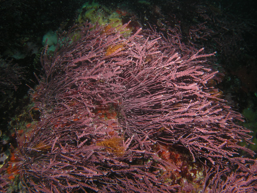
Rhodophyta (Red algae)"Rhodophyta (Red algae)" by Ed Bierman is licensed under CC BY 2.0

 Definition of Rhodophyta (Red Algae)

The term "Rhodophyta" comes from the Greek,  rhodos  which means "red". So, Rhodophyta means red algae ( red algae ). Unlike other phyla, this phylum does not have flagella stages in its life cycle. Members of this phylum have photosynthetic pigments in the form of phycobilins consisting of  phycoerythrin  (red pigment) and  phycocyanin  (blue pigment). In addition to these two pigments, Rhodophyta also has chlorophyll a and b and carotenoids.

Phycoerythrin is the most dominant pigment that causes the color of the thallus of this algae to turn red. However, not all of these algae are red. In the deep sea, this algae has a purple almost black color. At medium depths it is bright red, while at very shallow water, it is slightly greenish.

When this has been known for about 2,500 species, mostly live in the sea, especially the tropics and often called  kelp  ( sea weed ) because of her body shape like grass. Rhodophyta grows on rocks in tidal areas to depths of up to 90 meters below sea level where certain light waves from sunlight are still able to reach them.

Rhodophyta thallus is relatively large, but rarely exceeds 90 cm in length. Some species are filamentous, but most form complex structures that branch like feathers or are flattened and spread like ribbons. Generally, Rhodophyta are multicellular, but there are also unicellular Rhodophytes. Multicellular red algae are generally macroscopic and their body structure resembles a plant ( thallus ). Thallus in Rhodophyta in the form of strands or like plants.

Characteristics of Rhodophyta (Red Algae)

Red algae (Rhodophyta) have general characteristics or characteristics, including the following.

■ Core are eukaryotic cells because the cell nucleus already had membranes.  

■ Most multicellular (multicellular).  

■ Generally, macroscopic (visible with the naked eye) with a length of 1 m.  

■ The only algae that do not have flagella phase in its life cycle.  

■ Characteristically autotorof, because chlorophyll to perform photosynthesis.  

■ The chloroplasts contain pirenoid to store the results of photosynthesis.  

■ Backup food is stored in the form of flour fluoride (a type of carbohydrate), floridosid (compounds of glycerine and galactose) and drops of oil. Floridoside will have a reddish color when added with iodine.  

■ The shape of the talus in the form of a strand or shaped like a tree.  

The thallus is red to purple in color but some are blonde or reddish.  

■ His body was covered with calcium carbonate (CaCO 3 ).  

■ The cell walls consist of layered components. The inner cell wall is composed of myofibrils, while the outer cell is composed of mucus.  

■ Have pigment chlorophyll a and b, carotenoids, phycocyanin (blue) and the dominant pigment fikoeritrin (red).  

How to Reproduction Rhodophyta (Red Algae)

Red algae can reproduce asexually (vegetatively) and sexually (generatively). Asexual reproduction by forming aplanospores , namely nonmotile spores (not moving) and derived from diploid algae thallus. Furthermore, these spores will grow into new red algae. In Rhodophyta, fragmentation asexual reproduction is rare.

Sexual reproduction (generative) occurs by  oogamy , and in some species undergo alternation of offspring (metagenesis). Sexual reproduction is carried out by the fusion of male gametes that have no locomotion (sperm) and ova. The male gametes are formed in the spermatangium, while the female gametes are formed in the carpogonium. The next fertilized zygote will grow into a diploid red algae.

The following is a life cycle chart or alternation of offspring in an example of a red algae species, Polysiphonia.

Based on the picture above, try to write down the sequence of asexual (vegetative) reproduction starting from the sporophyte. Then, also write down the sequence of sexual reproduction (generative) to the sporophyte!

The sporophyte produces  meiospores  which will develop into gametophytes. The gametophyte forms  spermatangia  which produce  spermatia  and carpogonium  which contains  trichogen cells . The spermatia attach to the tip of the trichogen, continuing to enter the base of the cell. Here the fusion occurs between the nucleus of the sperm and the nucleus of the female cell to form a zygote (goninoblast).  Goninoblast  is a filament that is formed from the zygote and at the end of the filament a carposporangium is formed .

Furthermore, in the carposporangium formed carpospora.  Carpospora  out of the carposporangium, to further grow into a sporophyte (new Polysphonia). In its growth, Polysiphonia undergo alternation of offspring (metagenesis), namely asexual reproduction and sexual reproduction take place alternately.

Examples and Roles of Rhodophyta (Red Algae) in Life

Some red algae serve as important supports for tropical coral reefs. Red algae is an important food ingredient in Asian countries. In Japan, for example, red algae are dried and used in a number of dishes. In addition to producing algin, red algae also produce carrageenan and agar.

Carrageenan ( carrageenan ) is a type of polysaccharide used as an ingredient in cosmetics and gelatin capsules and is an additive that can be added to puddings and ice cream. To be used as food. Apart from being used as a food ingredient, agar-agar is also used as a medium for microorganism culture, cosmetics, medicine, canned meat coating, ice cream hardener, as well as fat emulsifier and chocolate bars.

Some examples of Rodophyta are as follows.

■ Eucheuma spinosum , widely cultivated because it produces so many are in Indonesian waters.  

■ Chondrus crispus , also cultivated known as seaweed.  

■ Gelidium coulteri  and  Gracilaria sp. , as an ingredient for making gelatin, is widely available in cold waters of the country.  

■ Carolina sp.  is a member of Rhodophyta (red algae) whose body is coated by calcium carbonate.  

■ Dasya, Batracnospermum, Scinaiafurcellata,  Porphyra perforated , Polysphonia,  Halosaccion glandiforme ,  Bossea orbigniana , and so on.

Phaeophyta (Brown Algae): Definition, Characteristics, Classification, Reproduction, Examples and Role for Life

Phaeophyta Fucus receptacle 03"Phaeophyta Fucus receptacle 03" by Bruce Kirchoff is licensed under CC BY 2.0

 Definition of Phaeophyta (Brown Algae)

Brown algae are plant-like protists that have many-celled thallus, so they can be seen macroscopically (with the naked eye). The thallus has an adhesive device to attach its body to the substrate, while the rest of the body floats on the water. Some members of the Phylum Phaeophyta such as Sargassum, Macrocystis, and Nereocystis have air bubbles that function to store nitrogen gas and to float on the surface of the water.

Brown algae contain brown pigments (xanthophylls), chlorophyll a and c. The amount of xanthophyll pigment is dominant, causing the color of the thallus to be brown. Food reserves are stored in the form of laminarin. Generally, brown algae are multicellular (multicellular). Its body shape resembles higher plants because it has parts resembling roots, stems, and leaves, making this algae easy to identify.

About 1,500 species of Phaeophyta or brown algae are known. Almost all types of Phaeophyta live in the sea, especially in cold areas, which live on rocks at the bottom of the water as deep as 1.5  -  5 meters from the water surface. All brown algae are threads or sheets and are autotrophic (capable of producing their own food).

All Phaeophyta live in colonies with shapes varying from simple to large ones with complex cell organization. In large colonies of Phaeophyta, no real organs have yet been formed, although in some species there are shapes resembling roots, stems, and leaves, the whole part is referred to as a thallus.

Characteristics of Phaeophyta (Brown Algae)

Brown algae (Phaeophyta) have general characteristics or characteristics, which are as follows.

■ Core are eukaryotic cells because the cell nucleus already had membranes.  

■ multicellular (multicellular).  

■ Shaped sheet, and some even resembling a tall plant (Plantae) because it has a section resembling roots, stems, and leaves.  

■ It has air bubbles that served as a float.  

■ Had the talus microscopic to macroscopic size.  

■ Have pigment chlorophyll a, chlorophyll c, violaxantin, b-carotene, diadinoxantin, as well as the amount xantofil dominant.  

In the form of branched filaments, unbranched and some are erect.  

■ Characteristically autotorof, because chlorophyll to perform photosynthesis.  

■ Having a single chloroplast some thread-like disk-shaped (discoid).  

■ The chloroplasts contain pirenois to store food reserves.  

■ Backup stored food in the form of laminarin.  

■ Have a cell wall.  

■ In the cell wall and intercellular space are agen (alginate acid), the inside of the cell wall composed of cellulose layers.  

■ Having a network of transportation such as higher plants.  

■ Almost all types of Phaeophyta habitat in the ocean, especially in the cold, that live in the rocks in the bottom waters as deep as 1,5  -  5 meters from the water's surface.  

■ All Phaeophyta live in colonies with varied shapes from the simple to the form of large (over 30 meters) with complex cellular organization.

Classification of Phaeophyta (Brown Algae)

■ Laminaria , has a stem, the leaf-shaped sheets, containing iodine and alginic acid.  

■ Macrocystis , produces iodine and alginic acid that serves as an industrial material.  

■ Sargasum , sheet-shaped leaves, between the stem and the stem there are air bubbles.  

■ Fucus , leaf shape in the form of sheets and at the edges of the leaves are bubbles.

Methods of Reproduction of Phaeophyta (Brown Algae)

Reproduction in Phaeophyta is done asexually (vegetatively) and sexually (generatively). Brown algae asexual reproduction is done by fragmentation  and  formation of spores  (aplanospores and zoospores). The resulting zoospores have flagella that are unequal in length and are located on the lateral side (sides or edges).

Meanwhile, sexual reproduction is done by  isogamy ,  anisogamy , or  oogamy . Fucus vesiculosis is an example of brown algae that reproduces oogamously. There is a way of generative/sexual reproduction of brown algae which is similar to higher plants, namely the ends of the fertile thallus sheets form a body containing reproductive organs called  receptacles .

Inside this receptacle there is a  conceptacle  containing antheridia which produces male sex cells in the form of spermatozoa and oogonia which produce eggs (ova) and sterile threads called paraphysis. The antheridia are funnel-shaped cells that arise from the base and edges of the conceptacle, the oogonia are bodies that sit on top of the stalk.

If a spermatozoon can fertilize an egg, a zygote will be formed. The zygote then forms a thick wall of cellulose and pectin, then attaches to a substrate such as rocks, then grows into a new individual whose chromosomes are diploid.

Examples and Roles of Phaeophyta (Brown Algae) in Life

Many types of Phaeophyta are beneficial to humans. Some species produce human food. In other countries kelp is used for animal feed and fertilizer, because the nitrogen and potassium content is high but the phosphorus content is low. Phaeophyta also produce algin (alginic acid), a colloid that is useful as a stabilizer in the manufacture of ice cream. Algin is also important in the pharmaceutical industry, namely as an ingredient for making pills, tablets, ointments, and dental cleaning drugs.

Some examples of Phaeophyta are as follows.

■ Fucus vesiculosus , height can reach 30  -  100 cm, live attached to rocks that appear when water is low. There are air bubbles along the sides of the thallus that branch like a fork. The tip is enlarged to form a conceptacle.  

■ Sargassum siliquosum , live attached to rocks along the rocky coast of the tropics. However, on the northern Atlantic coast, the species of  Sargasssum natans  floats freely on the sea surface. Sargassum sizes vary from small to hundreds of meters in length.  

■ Macrocystis integrifolia  or  kelp , size is very large, on the west coast of North America are found in length can reach three kilometers. Living kelp is firmly attached to rocks with the help of a root-like thallus.  

■ Laminaria sinclairii  is a type of brown algae producer alginic acid needed for the production of textiles, food, and cosmetics.  

■ Fucus serratus , including brown algae that differentiate into a form that floats.  

■ Postelsia  is an example that many common brown algae.  

■ Turbinaria decurens ,  Dictyota sp .,  Dictyosiphon sp .,  Nereocystis sp . is another example of Phaeophyta species or brown algae.